
Radiology and X-ray Equipment
Mammography: A radiograph of the breast that is used for cancer detection and diagnosis. Tumors tend to appear as regular or irregular-shaped masses that are somewhat brighter than the background on the radiograph (i.e., whiter on a black background or blacker on a white background). Mammograms can also detect tiny bits of calcium, called microcalcifications, which show up as very bright specks on a mammogram. While usually benign, specific patterns of microcalcifications could indicate the presence of cancer. Learn more about mammography
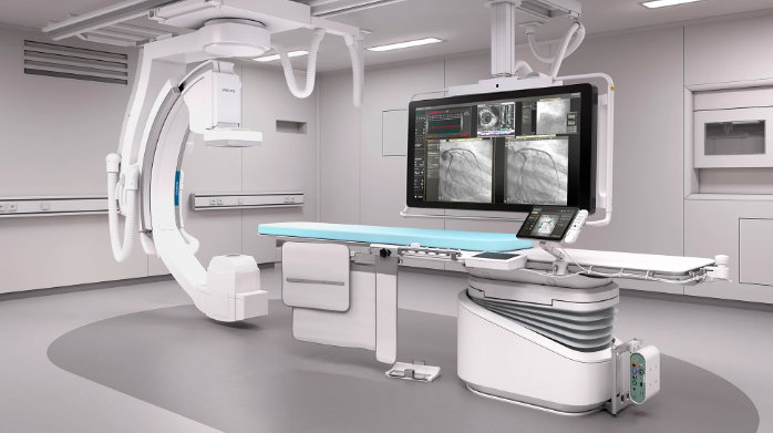
1. X-Ray Equipment
X-ray machines use radiation to produce images of the inside of the body. The basic components of an X-ray system include:
- X-ray Tube: The source of X-rays, it generates the radiation. The tube consists of a cathode (electron emitter) and an anode (target for electrons).
- Control Panel: Where radiologic technologists set the exposure factors like time, voltage (kV), and current (mA) for the X-ray.
- Detector/Film: The system captures the X-ray image. Traditional film has been largely replaced by digital detectors.
- Lead Aprons/Shields: Used for protection to minimize radiation exposure to parts of the body not being imaged.
- Collimator: A device that shapes and limits the X-ray beam to focus only on the area of interest.
- Radiographic Table: The table where the patient lies, or in some cases, sits, during the X-ray process.
- Image Receptors: Modern systems use digital receptors, including flat-panel detectors or computed radiography (CR) cassettes, to capture images.
2. Types of X-Ray Machines
- Conventional X-Ray: Standard X-ray for imaging bones, organs, and some soft tissues.
- Fluoroscopy: A continuous X-ray used to visualize movement in real-time (e.g., swallowing, blood flow).
- Mammography: Special X-ray for breast tissue.
- Dental X-rays: Compact X-ray machines designed for dental diagnostics.
3. Radiology Equipment Beyond X-Ray
Radiology includes many other imaging techniques beyond X-rays, such as:
- CT (Computed Tomography): Uses X-rays in a rotating motion to create detailed cross-sectional images of the body.
- MRI (Magnetic Resonance Imaging): Uses strong magnets and radio waves to produce detailed images of soft tissues.
- Ultrasound: Uses sound waves to generate images, typically for monitoring pregnancies or organs like the heart.
- Nuclear Medicine: Involves radioactive substances to visualize and treat conditions.
4. Advances in X-Ray Technology
- Digital Radiography (DR): Modern X-ray systems use digital sensors that capture images directly without the need for film processing. This leads to faster imaging and improved image quality.
- Portable X-Ray: Mobile systems allow for X-rays to be taken at the patient’s location (e.g., bedside or in emergency situations).
5. Safety Considerations
Radiology and X-ray use ionizing radiation, which can be harmful in large doses, so safety protocols are important:
- Minimizing Exposure: Use of protective equipment (like lead aprons) and limiting exposure to the necessary parts of the body.
- Radiation Dose Monitoring: Using the lowest effective dose of radiation for imaging.
6. Key Terms in Radiology

- Radiologist: A doctor specialized in interpreting medical images.
- Radiologic Technologist (RT): A healthcare professional trained to operate radiology equipment.
- Contrast Agents: Substances like iodine or barium that help enhance images by improving the visibility of organs or tissues during an X-ray.